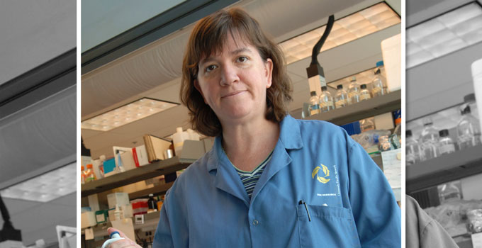
Animal models are often used to study how a cancer evolves or how effective a treatment could be, but Dr. Laurie Ailles, Scientist at the Princess Margaret Cancer Centre and OICR Investigator, has recently found that certain animal models could be used to predict the aggressiveness of a patient’s oral cancer and help inform treatment decisions in the clinic.
Ailles studies head and neck squamous cell carcinoma in mouse xenograft models – mice that are implanted with a human patient’s tumour tissue. She observed that some patients’ tumours could not grow in these xenograft models, where other patients’ cancers could grow and do so very quickly.
To investigate these irregular growth patterns, Ailles linked xenograft data with human patient clinical data and found a strong correlation between a patient’s survival and how their tumour could grow in mice. Her study, which was recently published in Cell Reports, identified that the rapid en grafters – the patients whose tumour tissue could grow rapidly in mice – had far worse survival outcomes than the patients whose tumours couldn’t grow in mice.
“We’ve been using xenograft models to study head and neck cancers for years,” says Ailles. “But this study reveals an interesting and important link between engraftment kinetics and patient survival, which is something we’ve never looked at before.”
Ailles’ study was the first to investigate the speed at which oral tumours grow in xenograft models. The research group found that in fewer than eight weeks, they could identify which models were rapid engrafters and thus which patients had more aggressive cancers. This eight-week timeframe could allow clinicians to use engraftment to guide treatment selection in the clinic.
“In the clinic we see that oral cancers – which have a five-year survival rate of only 50 per cent – grow differently in different patients and there’s no clearway to predict these outcomes,” says Ailles. “Our study shows that xenograft engraftment patterns are a strong predictor of disease outcomes and can be measured in a clinically relevant timeline.”
Ailles looks forward to performing broader genomic profiling and deeper analyses on the drivers and markers of engraftment. “If we can understand why some patients’ tumours are rapid engrafters and some aren’t, we could even find new ways to treat the disease.”
